
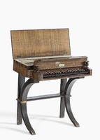
Glasschord, Heinrich M. Fuchs
Nuremberg, Germany ca. 1820, Inv. No. 45933
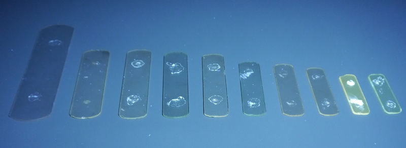
Yellowish fluorescence of the small glass plates (right), indicating the presence of manganese (acquired with a Hönle UVAHAND 250 GS handheld UV-lamp, Intensity 250 mW/cm2 UVA). (Attribution) Photo: Deutsches Museum, C. Holzer CC BY-SA 4.0
UV light fluorescence could be performed on a set of separate glass plates that are stored separately from the instrument. Under visible light, differences between glasses of colorless, green and pinkish shades, which are related to the ingredients and the condition of the glasses, were visible. The examination under UV-light confirmed the presence of manganese in the pinkish glass plate, because it showed a clear yellow fluorescence.
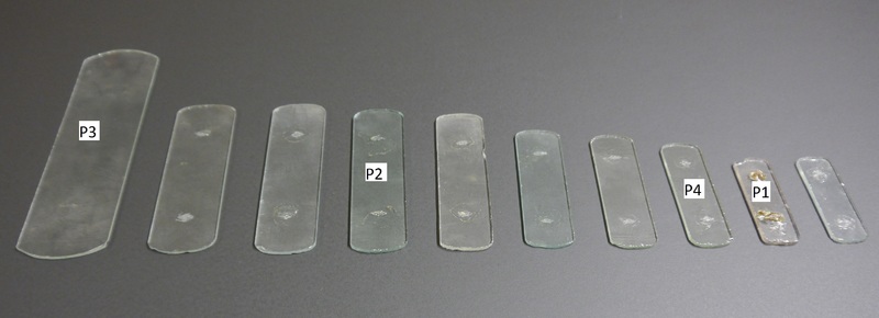
XRF measurement points on the separate glass plates. (Attribution) Photo: Deutsches Museum, C. Holzer CC BY-SA 4.0
XRF was conducted on the pinkish plate (P1), the greenish one (P2) and the two nearly colorless ones (P3, P4) in ambient conditions. Two spectra were taken from each plate in the same spot to detect both low- and heavy-weight elements.
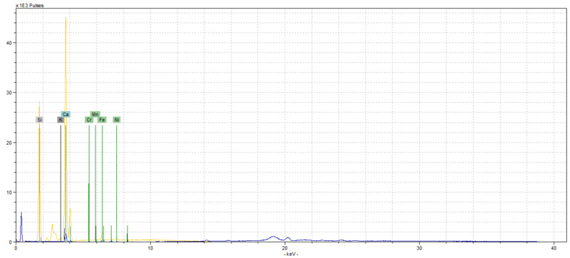
The glass plates were made of a potash-silica glass, which was stabilized with calcium oxide. In addition, arsenic or lead was added to the mixture, to improve the workability of the glass while retaining stability. The presence of manganese as a decolorizer, that was presumed after the UV-spectroscopy, could be confirmed. This was especially pronounced in the pinkish plate. Both iron and copper were detected in the greenish glass.
From a preservation perspective, the glasses are susceptible to high levels of humidity, because of the potash fluxing agent, and to long time exposure to UV light (several years). The high energy electromagnetic radiation can cause reverse oxidation in the manganese oxide, that was originally added as a decolorizer. In the oxidized state, this component turns the glass slightly pinkish or purplish. The effect can be seen on one of the glass plates from the glasschord.
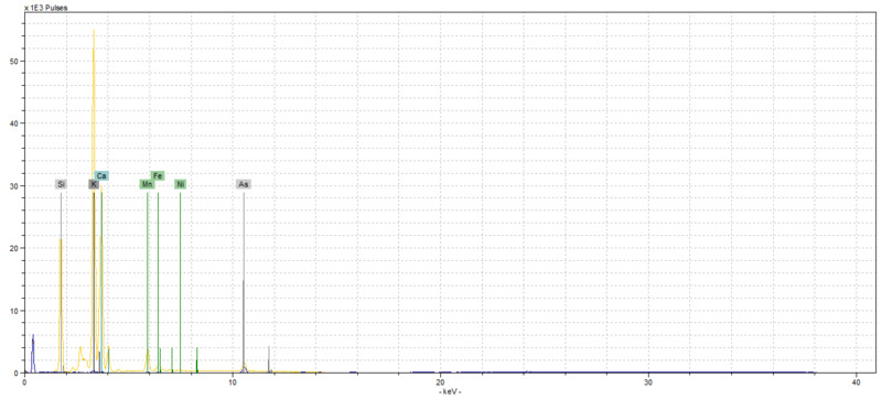
XRF spectrum of the small pink glass plate from the glasschord (Inv. No. 45933, P1) taken with a Bruker Titan S1 600-800 handheld X-Ray Fluorescence spectrometer with a Si-PIN detector (Hardware settings: Source: Rh; Phase 1: Voltage: 45 kV, Current: 12 µA,TiAl filter, Acquisition Time 30s; Phase 2: Voltage: 15 kV, Current: 29 µA, Acquisition Time 30s; Software: Bruker Artax Spectra). (Attribution) Photo: Deutsches Museum, C. Holzer CC BY-SA 4.0
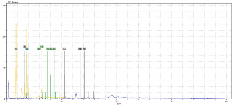
XRF spectrum of the small green glass plate from the glasschord (Inv. No. 45933, P2) taken with a Bruker Titan S1 600-800 handheld X-Ray Fluorescence spectrometer with a Si-PIN detector (Hardware settings: Source: Rh; Phase 1: Voltage: 45 kV, Current: 12 µA,TiAl filter, Acquisition Time 30s; Phase 2: Voltage: 15 kV, Current: 29 µA, Acquisition Time 30s; Software: Bruker Artax Spectra). (Attribution) Photo: Deutsches Museum, C. Holzer CC BY-SA 4.0
XRF spectrum of the large colorless glass plate from the glasschord (Inv. No. 45933, P3) taken with a Bruker Titan S1 600-800 handheld X-Ray Fluorescence spectrometer with a Si-PIN detector (Hardware settings: Source: Rh; Phase 1: Voltage: 45 kV, Current: 12 µA,TiAl filter, Acquisition Time 30s; Phase 2: Voltage: 15 kV, Current: 29 µA, Acquisition Time 30s; Software: Bruker Artax Spectra). (Attribution) Photo: Deutsches Museum, C. Holzer CC BY-SA 4.0

XRF spectrum of the small colorless glass plate from the glasschord (Inv. No. 45933, P4) taken with a Bruker Titan S1 600-800 handheld X-Ray Fluorescence spectrometer with a Si-PIN detector (Hardware settings: Source: Rh; Phase 1: Voltage: 45 kV, Current: 12 µA,TiAl filter, Acquisition Time 30s; Phase 2: Voltage: 15 kV, Current: 29 µA, Acquisition Time 30s; Software: Bruker Artax Spectra). (Attribution) Photo: Deutsches Museum, C. Holzer CC BY-SA 4.0
Click here for more information on the glasschord.
Citation: Charlotte Holzer, ‘Findings from Non-destructive Material Analysis on Musical Glass Instruments’, in: Materiality of Musical Instruments. A Virtual Exhibition.